More Information

Submitted: May 07, 2024 | Approved: May 14, 2024 | Published: May 15, 2024

How to cite this article: Ajao FO, Kalejaiye NO, Iyedupe MO, Abiodun S, Gbadero J, et al. Cardioprotective Potentials of Anacardium occidentale Nuts Methanolic Extract in Diabetes-Induced Cardiac Dysfunction in Rats. Arch Pharm Pharma Sci. 2024; 8: 056-066.

DOI: 10.29328/journal.apps.1001057

Copyright License: © 2024 Ajao FO, et al. This is an open access article distributed under the Creative Commons Attribution License, which permits unrestricted use, distribution, and reproduction inany medium, provided the original work is properly cited.

Keywords: Anacardium occidentale nuts; Diabetes mellitus; Oxidative stress & inflammation; Cardiac enzymes & lipid profile; Cardiac apoptosis

 FullText PDF

Cardioprotective Potentials of Anacardium occidentale Nuts Methanolic Extract in Diabetes-Induced Cardiac Dysfunction in Rats

Folasade Omobolanle Ajao1*, Noheem Olaoluwa Kalejaiye1, Marcus Olaoye Iyedupe1, Sunday Abiodun1, Joy Gbadero1, Pelumi Ogundele1, Zainab Adeagbo1, Oluwatosin Ojolo1, Enitan Shonde1 and Funmilayo Elizabeth Olaleye2

1Physiology Department, Faculty of Basic Medical Science, College of Health Science, Ladoke Akintola University of Technology, P.M.B. 4000, Ogbomoso, Oyo State, Nigeria
2Department of Human Nutrition and Dietetics, College of Medicine, University College Hospital, Ibadan, Oyo State, Nigeria

*Address for Correspondence: Folasade Omobolanle Ajao, Physiology Department, Faculty of Basic Medical Science, College of Health Science, Ladoke Akintola University of Technology, P.M.B. 4000, Ogbomoso, Oyo State, Nigeria, Email: foajao@lautech.edu.ng

Background: The unwanted adverse toxicity displayed by synthetic antidiabetic medicine leads to the search for effective natural medicine to combat diabetes complications. This study investigated the cardioprotective of Anacardium occidentale nuts methanolic in high-fat diet (HFD)/streptozotocin (STZ)-induced diabetic rats.

Materials and methods: Forty male adult Wistar were used and fed with HFD for 6 weeks before diabetes induction. The rats were grouped into 5 groups, 8 rats/group. Group I: normal control; Group II: diabetic control; Group III & IV: diabetic rats + 100 mg/kgb.wt & 200 mg/kgb.wt Anacardium occidentale nuts methanolic extract; Group V: diabetic rats + 200 mg/kgb.wt metformin. The rats were sacrificed on the experiment’s last day, blood samples were collected and the hearts were isolated for biochemical parameters estimation.

Results: Food intake, water intake, plasmas insulin, Fasting Blood Glucose (FBG), glycosylated hemoglobin (HbA1c), cardiac enzymes, lipid profile, inflammatory cytokines, malondialdehyde, fibrotic marker, caspase-3 in cardiac of diabetic rats were elevated (p < 0.05) significantly. Body weight, cardiac antioxidant, and anti-apoptotic marker levels diminished (p < 0.05) significantly in diabetic rats. 100 mg/kgb.wt & 200 mg/kgb.wt of Anacardium occidentale nuts methanolic extract administration significantly suppressed the plasma insulin, FBG, HbA1c, cardiac lipid profile, cardiac enzymes biomarker, cardiac inflammatory cytokines, cardiac malondialdehyde, cardiac fibrotic marker, cardiac caspase-3, food intake & water intake and increased the body weight, cardiac antioxidant & cardiac anti-apoptotic marker in the diabetic rats.

Conclusion: Anacardium occidentale nuts attenuate cardiac injury in diabetes. It could be a natural medicine to manage diabetes-cardiovascular complications.

The prevalence of diabetes mellitus escalates significantly [1]. According to the International Diabetes Federation, it is projected that diabetes will affect approximately 783.2 million individuals by 2045 [2].

Diabetes mellitus is described as a chronic metabolic disorder due to impairments in pancreatic β-cells insulin secretion, insulin action, or both which disrupt the metabolism of carbohydrates, lipids, and proteins, leading to chronic hyperglycemia [3-5]. Noticeably, untreated sustained hyperglycemia is implicated in the pathogenesis of diabetes-related macro-vascular and micro-vascular complications, including nephropathy, neuropathy, retinopathy, and cardiovascular diseases [6].

Cardiovascular complications are recognized as the foremost cause of mortality and morbidity in type 1 and type 2 diabetic patients [7]. Cardiovascular complications cause 80% of mortality in diabetes conditions [8].

Diabetes treatment strategies in recent decades have advanced. Nevertheless, anti-diabetic medications have severe side effects [9]. The World Health Organization (WHO) has shifted attention to the use of medicinal plants to manage diabetes mellitus [10,11]. Medicinal plants possess notable anti-diabetic compounds including flavonoids, alkaloids, phenolics, and tannins with few or no adverse effects [12].

Anacardium occidentale Linn (A. occidentale) globally known as the cashew tree is a member of the Anacardiaceae family and is grown widely in tropical countries. A. occidentale has been used as a folk remedy for treating a range of diseases including diabetes mellitus [13]. The species of this plant possess abundant phenolic compounds and flavonoids in their leaves, bark, fruits, and nuts. These compounds exhibit potent anti-inflammatory and antioxidant properties, providing cellular protection [14]. Anti-inflammatory, anti-oxidative, and analgesic activities of the nuts have been previously reported [15]. Also, in mild hyperhomocysteinemia rats, oral administration of A. occidentale nuts was reported to counteract biochemical changes, oxidative stress, pro-inflammatory cytokine release, histological tissue injuries, fibrosis, and apoptosis in the kidney, colon, and liver [16]. However, research on A. occidentale nuts to protect and

manage cardiac complications in diabetes has never been elucidated. This recent study scientifically investigated the potential of A. occidentale nuts methanolic extract to attenuate cardiac injury in high-fat diet/streptozotocin-induced diabetic rats.

Chemicals and Drugs: Streptozotocin, methanol, glucose, phosphate buffer, ketamine, and xylazine.

Experimental animals

Forty adult Wistar rats weighing (250 g – 300 g) were purchased from the Animal Research House of the Physiology Department, Ladoke Akintola University of Technology (LAUTECH), Ogbomoso, Oyo State, Nigeria. The animals were kept in a cleaned propylene cage and fed with standard pelletized feed with water ad libitum for 7 days to acclimatize under the pathogen-free environmental conditions of temperature (25 ± 2 °C), relative humidity (45% ± 5%) and 12:12 hour’s light/dark cycles. All experimental procedures were conducted according to the National Institutes of Health (NIH) Guide for the Care and Use of Laboratory Animals protocol and approved by the Faculty of Basic Medical Science Ethics Research Committee Ladoke Akintola University of Technology (Ethical Approval Number: ERCFBMSLAUTECH:021/01/2024).

A. occidentale nuts collection and extraction

A. occidentale plant was identified at LAUTECH Agriculture Research Farm, authenticated, and assigned a voucher number LH0533 by Dr. A. T. J. Ogunkunle of the Biology Department, LAUTECH. The freshly plunged A. occidentale nuts were washed thoroughly with distilled water, air-dried, and removed from the outer coat before extraction. The nuts were grinded to powder form, kept in an air-tight container, and 500 g of the nut’s powdered form was extracted with 95% methanol in a Soxhlet apparatus to form a semi-solid. The semi-solid was then evaporated in a rotary evaporated pressure and the solid formed was collected and stored at -4 °C until needed.

Diabetes induction

The animals were fed with a High-Fat Diet (HFD) for 6 weeks before diabetes induction. Then, subjected to overnight fasting (12 hours) before diabetes. The animals were injected intraperitoneally with a repeated single dose of freshly prepared streptozotocin (35 mg/kgb.wt) to induce diabetes and given a 2% glucose solution to prevent drug-induced hypoglycemic death. Diabetes induction was authenticated after 72 hours of streptozotocin injection via the animals’ tail prick venous blood using a glucometer (Accu-check) and test stripes. Animals with fasting blood glucose ≥ 200 mg/dL were confirmed diabetic and picked for the experimental study.

Experimental animal grouping

Forty rats were grouped into five groups, 8rats/group. Group I served as control rats, and Group II-V were HFD/STZ-induced diabetic rats treated with different doses of A. occidentale nuts methanolic extract as follows:

Group I: Normal control

Group II: Diabetic control

Group III: Diabetic + 100 mg/kgb.wt AONM Extract (Low dose)

Group IV: Diabetic + 200 mg/kgb.wt AONM Extract (high dose)

Group V: Diabetic + 200 mg/kgb.wt Metformin (reference drug)

A. occidentale nuts methanolic extract was administered for consecutive 6 weeks. Food intake, water intake, and body weight were recorded daily with weighing balance. Weekly fasting blood glucose levels were measured using the glucose-oxidase/peroxidase (GOD-POD) method via the pricked tail vein blood with a digital Accu-Chek glucometer and test strips and recorded throughout the treatment period of the experiment.

Blood Pressure and Electrocardiographic (ECG) parameters recording

The mean Systolic Blood Pressure (SBP) and Diastolic Blood Pressure (DSP) were measured using a non-invasive tail-cuffed method.

ECG was recorded using a three-lead non-invasive with electrodes positioned in lead II and sampled at 1 kHz. RR interval, PR interval, P-wave, QRS complex, QT interval, and heart rate were measured. QT was corrected from the QR interval using the Bazett formula [17]:

QTc = QT ( RR/f ) 1/2 where f = 150 ms

Biochemical assay

At the end of the treatment period, the rats were fasted overnight after the last A. occidentale nuts methanolic extract low and high doses (100 mg/kgb.wt & 200 mg/kgb.wt) were administered. The animals were anesthetized with ketamine (40 mg/kgb.wt) and xylazine (20 mg/kgb.wt) and sacrificed by cervical dislocation. Fasting blood samples were collected from the apex beat of the rats’ hearts and the hearts were isolated immediately after blood collection, rinsed in normal saline, and homogenized with freshly prepared cold phosphate-buffered. The blood samples were centrifuged at 3,500 rpm for 15 minutes at 4 °C and the heart tissues homogenates were centrifuged at 10000 rpm for 10 minutes at 4 °C. After centrifugation, the clear supernatant plasma obtained was used for biochemical parameters determination.

Glycated hemoglobin (HbA1c) was estimated using a rat hemoglobin HbA1c assay kit following the manufacturer’s instructions.

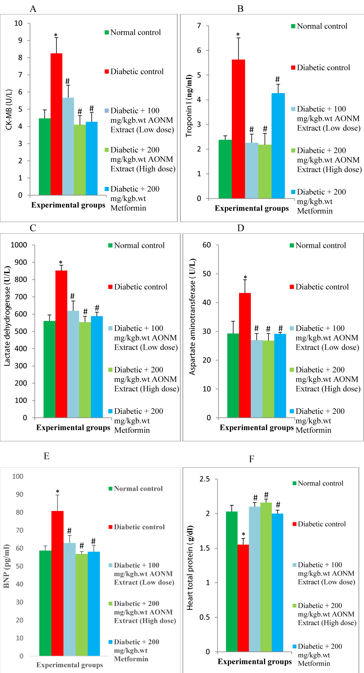
Enzyme-Linked Immunosorbent Assay (ELISA) was utilized to measure the levels of insulin, total protein, Creatine Kinase-Myocardial Band (CK-MB), cardiac troponin I, Interleukin-1β (IL-1β), Interleukin-6 (IL-6), Tumor Necrosis Factor-alpha (TNF-α), Brain Natriuretic Peptide (BNP), Transforming Growth Factor-β1 (TGF-β1), B-cell lymphoma-2 (Bcl-2) and caspase-3 in rats. Each assay employed a specific ELISA kit designed for rats, following the manufacturer’s instructions.

Lactate Dehydrogenase (LDH) and aspartate aminotransferase (AST) were determined using a spectrophotometer and assay method with an available commercial kit.

Cardiac lipid profile including Total Cholesterol (TC), Triglycerides (TG), and High-Density Lipoprotein cholesterol (HDL-C), was determined using enzymatic colorimetric methods with commercially available assay kits, following the manufacturer’s protocol. Low-Density Lipoprotein Cholesterol (LDL-C) was calculated using the Friedewald equation: LDL-C = TC - (HDL-C + TG/5) [18]. Cardiovascular Risk Indices (CRI) were calculated as the ratio of TG to HDL-C.

The Atherogenic Coefficient (AC) and Castelli’s Risk Index-1 (CRI-1) were computed using the following formulas:

Atherogenic Coefficient (AC) = (TC - HDL-C)/HDL-C.

Castelli’s Risk Index-1 (CRI-1) = TC/HDL-C.

To determine the cardiac oxidative stress Malondialdehyde (MDA) and antioxidants’ Catalase (CAT) and Superoxide Dismutase (SOD) activities, ELISA assay kits were employed as per the manufacturer’s guidelines.

Statistical analysis

Data were presented as the standard error of means (Mean ± SEM) and were analyzed with a statistical package for social science (SPSS version 21.0 software) using one-way analysis of variance (ANOVA) followed by Tukey’s posthoc test to determine the statistically significant difference between groups. Data at p < 0.05 was considered statistically significant.

Effect of A. occidentale nuts methanolic extract on body weight and relative heart weight in HFD/STZ-induced diabetic rats

Diabetic-induced rats displayed a significant (p < 0.05) reduction in body and relative organ weights compared with normal control rats. The administration of 100 mg/kgb.wt (low dose) and 200 mg/kgb.wt (high dose) A. occidentale nuts methanolic extract improved the body and relative organ weights compared with diabetic control rats (Table 1).

Effect of A. occidentale nuts methanolic extract on food and water intake in HFD/STZ-induced diabetic rats

Food and water intake in HFD/STZ-induced diabetic rats increased (p < 0.05) significantly in comparison with normal control rats. The low dose (100 mg/kgb.wt) and high dose (200 mg/kgb.wt) A. occidentale nuts methanolic extract administration reduced the food and water intake compared with diabetic control rats (Table 1).

Effect of A. occidentale nuts methanolic extract on electrographic and blood pressure in HFD/STZ-induced diabetic rats

The ECG recording of the diabetic rats showed significant (p < 0.05) elevated, P-wave, P-R interval, Q-T interval, and QTc compared with the control. QRS complex, and heart rate (HR) diminished (p < 0.05) significantly in diabetic rats compared with the control rats. Administration of 100 mg/kgb.wt (low dose) and 200 mg/kgb.wt (high dose) A. occidentale nuts methanolic extract reversed the P-wave, P-R interval, Q-T interval, and QTc, and improved the QRS complex and HR in comparison with diabetic rats (Table 1).

The systolic blood pressure of diabetic rats increased (p < 0.05) significantly and diastolic blood pressure decreased significantly in comparison with control rats. Treatment with 100 mg/kgb.wt (low dose) and 200 mg/kgb.wt (high dose) A. occidentale nuts methanolic extract significantly decreased the systolic and diastolic blood pressure compared with the diabetic rats (Table 1).

Effect of A. occidentale nuts methanolic extract on plasma insulin, fasting blood glucose and glycosylated hemoglobin in HFD/STZ-induced diabetic rats

The level of insulin, fasting blood glucose, and glycosylated hemoglobin in diabetic-induced rats were significantly (p < 0.05) higher compared with normal control rats. The low dose (100 mg/kgb.wt) and high dose (200 mg/kgb.wt) A. occidentale nuts methanolic extract administration lowered the insulin, fasting blood glucose, and glycosylated hemoglobin in comparison with diabetic control rats (Figure 1A-1C).


Download Image

Figure 1: Effect of A. occidentale nuts methanolic extract low and high dose on (a) plasma insulin (b) fasting blood glucose (c) glycosylated hemoglobin in HFD/STZ-induced diabetic rats. Values are expressed as mean ± SEM (n = 8). *significant at p < 0.05 compared with control; #significant at p <0.05 compared with untreated diabetic group.

Effect of A. occidentale nuts methanolic extract on cardiac biomarkers’ and total protein in HFD/STZ-induced diabetic rats

HFD/STZ-induced diabetic rats demonstrated a significant (p < 0.05) rise in levels of cardiac biomarkers’ Creatine-Kinase Myoglobin (CK-MB), troponin I (TnP-I), Lactate Dehydrogenase (LDH), aspartate Aminotransferase (AST), Brain Natriuretic-Peptide (BNP), and a significant (p < 0.05) reduction in cardiac total protein level in comparison with normal control rats. A. occidentale nuts methanolic extract low dose (100 mg/kgb.wt) and high dose (200 mg/kgb.wt) administration remarkably diminished the cardiac CK-MB, TnP-I, LDH, AST, and BNP, and increased cardiac protein compared to diabetic control rats (Figure 2A-2F).


Download Image

Figure 2: Effect of A. occidentale nuts methanolic extract low and high dose on (a) creatine kinase-myocardial band (b) troponin I (c) lactate dehydrogenase (d) cardiac aspartate aminotransferase (e) brain natriuretic-peptide (f) cardiac total protein in HFD/STZ-induced diabetic rats. Values are expressed as mean ± SEM (n = 8). *significant at p < 0.05 compared with control; #significant at p < 0.05 compared with untreated diabetic group.

Effect of A. occidentale nuts methanolic extract on cardiac lipid profile, atherogenic coefficient and Castelli’s risk index-1 in HFD/STZ-induced diabetic rats

Cardiac triglyceride (TG), total cholesterol (TC), low-density lipoprotein-cholesterol (LDL-C), triglyceride/high-density lipoprotein-cholesterol ratio (TG/HDL-C ratio), atherogenic coefficient and Castelli’s risk index-1 significantly (p < 0.05) elevated, and high-density lipoprotein-cholesterol (HDL-C) significant (p < 0.05) diminished in HFD/STZ-induced diabetic rats compared with normal control rats. Administration of 100 mg/kgb.wt (low dose) and 200 mg/kgb.wt (high dose) A. occidentale nuts methanolic extract lowered the cardiac TG, TC, LDL-C, TG/HDL-C ratio, atherogenic coefficient, and Castelli’s risk index-1, and elevated the HDL-C in comparison with diabetic control rats (Table 2).

Table 1: Effect of A. occidentale Nuts Methanolic Extract Low and High Dose on Body Weight, Relative Heart Weight, Food Intake, Water Intake, Blood Pressure and Electrocardiographic Parameters in HFD/STZ-Induced Diabetic Rats.
 

Parameters

Experimental groups
Normal control Diabetic control Diabetic + 100 mg/kgb.wt AONM Extract (Low dose) Diabetic + 200 mg/kgb.wt AONM Extract (High dose) Diabetic + 200 mg/kgb.wt Metformin
Body weight (g) 262.20 ± 4.10 168.20 ± 8.40* 206 ± 7.10# 229.60 ± 11.10# 263.33 ± 6.96#
Relative heart weight (g) 0.01 ± 0.00 0.00 ± 0.00* 0.01 ± 0.00# 0.01 ± 0.00# 0.01 ± 0.00#
Food intake (g/day/rat) 23.52 ± 1.14 24.86 ± 0.89* 23. 42 ± 1.62# 25.31 ± 1.25# 24.68 ± 2.49#
Water intake (ml/day/rat) 76.90 ± 3.96 95.14 ± 2.91* 82.03 ±3.48# 76.57 ± 3.67# 75.43 ± 5.26#
SBP (mmHg) 137 ± 7.5 149 ± 15.03* 146 ± 12.9# 140 ± 8.29# 126 ± 6.02#
DBP (mmHg) 115 ± 9.84 113 ± 11.09* 103 ± 10.40# 92.3 ± 11.05# 90 ±5.19#
Heart rate (bpm) 256 ± 4.83 249 ± 10.11* 245 ± 0.41# 220 ± 3.64# 205 ± 3.87#
P-wave 22.5 ± 0.95 44.25 ± 2.32* 37.25 ± 0.85# 26.25 ± 1.70# 28.25 ± 1.18#
P-R interval 50 ± 1.47 65.75 ± 2.25* 57.5 ± 1.66# 54.25 ± 4.25# 53.75 ± 2.87#
Q-T interval 65.25 ± 2.25 145 ± 4.49* 96 ± 4.10# 88.75 ± 5.48# 79 ± 1.63#
QTc 134 ± 4.49 239. 25 ± 22* 183.5 ± 6.96# 173 ± 5.57# 145.25 ± 3.71#
QRS complex 13.25 ± 0.85 12.75 ± 0.85* 13.5 ± 0.64# 14.75 ± 0.25# 15 ± 0.82#
Values are expressed as mean ± SEM (n = 8). *significant at p < 0.05 compared with control; #significant at p < 0.05 compared with untreated diabetic group
Table 2: Effect of A. occidentale Nuts Methanolic Extract Low and High Dose on Cardiac Lipid Profile, Oxidative Stress Markers, and Antioxidants in HFD/STZ-Induced Diabetic Rats.
  Parameters Experimental groups
Normal control Diabetic control Diabetic + 100 mg/kgb.wt AONM Extract (Low dose) Diabetic + 200 mg/kgb.wt AONM Extract (High dose) Diabetic + 200 mg/kgb.wt Metformin
Heart TG (mg/dL) 110.78 ± 7.78 248.10 ±11.24* 115.96 ± 16.98# 99.91 ± 7.26# 104.09 ± 19.81#
Heart TC (mg/dL) 140.87 ± 9.73 230.75 ±16.93* 111.83 ± 30.58# 116.88 ± 7.72# 73.58 ± 30.86#
Heart LDL-C (mg/dL) 56.69 ± 5.94 146.45 ±17.77* 58.17 ± 9.19# 54.60 ± 10.55# 51.45 ± 4.55#
Heart HDL-C (mg/dL) 58.03 ± 1.21 34.68 ± 1.82* 50.19 ± 2.50# 46.30 ± 11.88# 36.22 ± 16.32#
TG/HDL-C ratio (mg/dL) 1.92 ± 0.15 7.19 ± 0.30* 2.34 ± 0.44# 1.64 ± 0.14# 1.84 ± 0.42#
Atherogenic Coefficient 1.42 ± 0.13 5.76 ± 0.69* 1.79 ± 0.39# 1.10 ± 0.27# 1.12 ± 0.20#
Castelli’s Risk Index 1 2.42 ± 0.13 6.76 ± 0.69* 2.79 ± 0.39# 2.10 ± 0.27# 2.12 ± 0.20#
Heart MDA (µM) 0.73 ± 0.03 1.85 ± 0.05* 0.84 ± 0.06# 0.89 ± 0.03# 0.87 ± 0.02#
Heart SOD (u/ml) 1.36 ± 0.03 0.60 ± 0.07* 1.39 ± 0.08# 1.48 ± 0.03# 1.44 ± 0.05#
Heart CAT (u/mg/protein) 24.55 ± 1.29 16.97 ± 0.53* 19.92 ± 1.65# 22.18 ± 1.56# 23.87 ± 1.45#
Values are expressed as mean ± SEM (n = 8). *significant at p < 0.05 compared with control; #significant at p < 0.05 compared with untreated diabetic group.
Effect of A. occidentale nuts methanolic extract on cardiac oxidative stress markers and antioxidants in HFD/STZ-induced diabetic rats

Antioxidants’ Superoxide Dismutase (SOD) and Catalase (CAT) levels in cardiac of diabetic-induced rats reduced (p < 0.05) significantly and oxidative stress markers’ Malondialdehyde (MDA) increased (p < 0.05) significantly in comparison with the normal control. The low dose (100 mg/kgb.wt) and high dose (100 mg/kgb.wt) A. occidentale nuts methanolic extract administration increased the cardiac SOD and CAT and reduced MDA when compared with diabetic control rats (Table 2).

Effect of A. occidentale nuts methanolic extract on cardiac inflammatory markers, apoptotic and anti-apoptotic markers in HFD/STZ-induced diabetic rats

There was a significant (p < 0.05) increase in cardiac interleukine-6 (IL-6), interleukine-1beta (IL-1β), tumor necrosis factor-alpha (TNF-α), transforming growth factor-beta1 (TGF-β1) in diabetic-induced rats compared with normal control rats. The administration of 100 mg/kgb.wt (low dose) and 200 mg/kgb.wt (high dose) A. occidentale nuts methanolic extract decreased the level of cardiac IL-6, IL-1β, TNF-α, and TGF-β1 in comparison with diabetic control rats (Table 3).

Table 3: Effect of A. occidentale Nuts Methanolic Extract Low and high Dose on Cardiac Inflammatory Markers, Apoptotic and Anti-Apoptotic Markers in HFD/STZ-Induced Diabetic Rats.
  Parameters Experimental groups
Normal control Diabetic control Diabetic + 100 mg/kgb.wt AONM Extract (Low dose) Diabetic + 200 mg/kgb.wt AONM Extract (High dose) Diabetic + 200 mg/kgb.wt Metformin
IL-1β (pg/ml) 6.75 ± 0.93 16.05 ± 2.13* 8.77 ± 0.81# 7.91 ± 0.42# 6.82 ± 1.58#
IL-6 (pg/ml) 49.57 ± 4.38 65.90 ± 5.07* 49.62 ± 2.21# 46.11 ± 2.94# 47.47 ± 2.27#
TNF-α (pg/ml) 16.80 ± 0.89 22.82 ± 1.78* 19.15 ± 1.41# 14.92 ± 1.42 16.68 ± 0.89#
TGF-1β (ng/ml) 0.29 ± 0.02 5.51 ± 0.29* 0.26 ± 0.02# 0.21 ± 0.01# 0.22 ± 0.00#
Bcl-2 5.96 ± 1.76 1.46 ± 0.37* 3.14 ± 0.39# 5.14 ± 0.44# 8.29 ± 0.42#
Caspase-3 7.25 ± 0.23 13.29 ± 0.6* 6.23 ± 0.61# 5.57 ± 0.48# 5.78 ± 0.29#
Values are expressed as mean ± SEM (n = 8). *significant at p < 0.05 compared with control; #significant at p < 0.05 compared with untreated diabetic group.

Cardiac apoptotic marker caspase-3 levels increased (p < 0.05) significantly and anti-apoptotic marker B-cell lymphoma-2 (Bcl-2) lowered (p < 0.05) significantly in cardiac diabetic rats when compared with the control. The low dose (100 mg/kgb.wt) and high dose (100 mg/kgb.wt) A. occidentale nuts methanolic extract administration increased the cardiac level anti-apoptotic Bcl-2 and reduced the apoptotic marker caspase-3 in comparison with the diabetic rats (Table 3).

Diabetes mellitus, an incurable chronic metabolic disease needs ample care for its management. Prolonged hyperglycemia causes many complications related to diabetic mellitus [19]. Cardiovascular ailment is one of the common diabetic complications. Oxidative stress, inflammation, and cell apoptotic are considerably implicated in cardiovascular disease manifestation in diabetes [20]. However, the World Health Organization data revealed that about twenty thousand medicinal plants are available worldwide [21,22]. Natural compounds in medicinal plants display strong pharmacological efficacy for managing many diseases. This study investigates the potential of A. occidentale nuts methanolic extract to attenuate cardiac damage in diabetes.

Diabetes animal models typically displayed clinical symptoms and features noticeable in human diabetes including, elevated blood glucose, decreased plasma insulin, dyslipidemia, frequent urination, polyphagia polydipsia, and loss of body weight [23]. Consistent with Raish, et al. [24] findings, the current experimental streptozotocin-induced diabetic rats developed hyperglycemia, hyperinsulinemia, polyphagia, polydipsia, and a reduction in body weight. Body weight loss in diabetes has been hypothesized to result from metabolic alterations that lead to excessive structural protein catabolism and reduce protein synthesis [25]. Excessive hepatic glycogenolysis and gluconeogenesis and decreased utilization of insulin by peripheral organs lead to hyperglycemia and hyperinsulinemia in diabetes [26]. Obviously, low (100 mg/kgb.wt) and high (200 mg/kgb.wt) dose A. occidentale nuts methanolic extract supplement ameliorates the aforementioned clinical features and symptoms in diabetic rats. Inhibition of hepatic gluconeogenesis, increase in hepatic glycogen storage and stimulation of peripheral tissues sensitivity to insulin action for glucose uptake could be likely mechanisms for the hypoglycemic and insulin-normalizing effects of the A. occidentale nuts.

As reported in various findings, hyperglycemia causes glycosylation of proteins which progresses to the formation of chronic elevated glycated hemoglobin HbA1c, a maker of glycemic control in diabetes [27]. This finding observed elevated HbA1c levels in diabetic rats, supports the other report. However, administration of low (100 mg/kgb.wt) and high (200 mg/kgb.wt) dose A. occidentale nuts methanolic extract suppressed the HbA1c levels and this might stem from the reduction in blood glucose level upon administration of the A. occidentale nuts extract, parallel the findings of Gothandam, et al. [28].

Diabetes patients frequently encounter alterations in the normal electrocardiography (ECG) pattern mostly the Q-T interval and T-wave. Diabetes specifically leads to prolongation of the QRS and Q-T intervals [29]. QTc stands for corrected Q-T interval and a remarkable prolongation of the Q-T interval serves as a crucial indicator for screening diabetic patients susceptible to sudden cardiac death [30,31]. This study observed bradycardia, prolonged P-wave, QT interval, and QTc intervals, and decreased QRS complex in the ECG pattern of diabetic rats. Moreover, supplements of A. occidentale nuts methanolic extract low (100 mg/kgb.wt) and high (200 mg/kgb.wt) doses to the diabetic rats restored the normal ECG pattern. Many studies have reported the significance of bioactive compounds of medicinal plants in restoring and enhancing cardiac function in diabetic rats via normalizing cardiac electrical activities [32]. This improvement in cardiac ECG patterns noticed in diabetic rats could be attributed to the antioxidants from bioactive compounds of the A. occidentale nuts.

Also, abnormal Systolic Blood Pressure (SBP) and Diastolic Blood Pressure (DBP) in diabetic rats have been established [33]. Dysfunction in these parameters was obvious in this study as the diabetic rats exhibited high SBP and reduced DBP, contrary to the findings of Bulani, et al. [34]. A. occidentale nuts methanolic extract low (100 mg/kgb.wt) and high (200 mg/kgb.wt) dose administered to the diabetic rats ameliorates the altered blood pressure and this showed the nuts inhibit the progression of blood pressure impairment in diabetic rats.

Cardiac enzymes are key sensitive biomarkers for cardiac injury in a diabetic heart. These enzymes are abundant in cardiac muscle tissue and damage to this muscle evokes the release of the enzymes into the circulation [35]. Chronic hyperglycemia-induced oxidative stress causes injury to the cardiac muscle cells leading to the release of these enzymes into the blood [36]. Also, studies have reported the release of high Brain Natriuretic Peptide (BNP), a ventricular dysfunction biomarker in diabetic heart failure [37]. Elevated levels of these biomarkers have been previously reported in the hearts of diabetic rats [38], the finding of this study is in line with the report, as the diabetic rats exhibited elevated cardiac enzymes Creatine Kinase-Myocardial Band (CK-MB), troponin- I (cTnP-I), Lactate Dehydrogenase (LDH) and aspartate aminotransferase (AST) and similar with Saklan et al findings [39], overexpression of ventricular dysfunction biomarker BNP was observed. The low (100 mg/kgb.wt) and high (200 mg/kgb.wt) dose A. occidentale nuts methanolic extract administration lessened the cardiac enzymes biomarker, indicating cardiomyocyte cells protection of the nuts which might stem from the antioxidant potentials of A. occidentale nuts which harmonized the findings of Adoga, et al. [40].

Dyslipidemia in chronic hyperglycemia is considered to contribute to the pathogenesis of ischemic heart disease and the progression of heart failure by enhancing lipid toxicity and reactive oxygen species production in diabetic individuals [41]. Our results revealed dyslipidemia in the diabetic rats noticed by a high level of cardiac triglycerides (TG), Total Cholesterol (TC) low-density lipoprotein-cholesterol (LDL-C), atherogenic coefficient (AI), and Castelli’s risk index-1 (CRI-1) with low cardiac high-density lipoprotein-cholesterol (HDL-C), consistent with findings of Tangvarasittichai [42]. The levels of cardiac TG, TC, LDL, AI, and CRI-1 were reduced and HDL-C improved on treatment with low (100 mg/kgb.wt) and high (200 mg/kgb.wt) dose A. occidentale nuts methanolic extract, suggesting anti-hyperlipidemic and cardioprotective of the nuts and could be due to the suppression and inhibition of intestinal and hepatic acyl-coenzyme A (CoA) activity and this parallel with the reports of Yang, et al. [43].

Oxidative stress is well-known to trigger the mechanism that leads to myocardial contractility loss, cardiac fibrosis, cardiac inflammation, apoptosis of cardiac cells, and DNA damage via excessive generation of reactive oxygen species in cardiac tissues [44-47]. The heart is vulnerable to oxidative stress injury due to the low antioxidant enzymes capacity to scavenge free radicals [48]. Excessive cardiac oxidative stress and low antioxidant enzymes have been reported in the diabetic heart [49]. According to the previous report, elevated cardiac oxidative stress marker malondialdehyde (MDA) and low levels of antioxidants superoxide dismutase (SOD) and catalase (CAT) were observed in the diabetic rats of the current study. Administration of low (100 mg/kgb.wt) and high (200 mg/kgb.wt) dose A. occidentale nuts methanolic extract raised the cardiac endogenous antioxidants SOD and CAT and depressed the oxidative stress marker, this indicates cardiac oxidative stress attenuation with potent antioxidants properties of the nuts via the numerous bioactive compounds present in the nuts, which is in line with the findings of Li, et al. [50].

Oxidative stress-induced cardiac inflammation has been implicated in the development and progression of cardiac injury in diabetic patients [51]. Excessive production of myocardial pro-inflammatory cytokines tumor necrosis factor-alpha (TNF-α), interleukine-1β (IL-1β), and interleukin-6 (IL-6) have been reported in the etiology of cardiac function anomalies and fibrosis [52]. In support of the findings of Liang, et al. [53], up-regulation of cardiac cytokines TNF-α, IL-1β, and IL-6 are observed in the heart of diabetic rats of the current study. However, these up-regulated cytokines in the diabetic heart were suppressed with low (100 mg/kgb.wt) and high (200 mg/kgb.wt) dose A. occidentale nuts methanolic extract treatment, which evident cardiac anti-inflammatory properties of A. occidentale nuts and this may be attributed to the antioxidant efficacy displayed by suppressing the hyperglycemia induced-oxidative stress in the didactic heart, similar with the report of Arjumand, et al. [54].

Cardiac inflammatory cytokines and reactive oxygen species, the consequences of oxidative stress are crucial mechanisms in the pathogenesis of cardiac fibrosis through superfluous cardiac transforming growth factor-beta 1(TGF-β1) [55]. Noticeably in the current study, a high level of TGF-β1 was expressed in the cardiac of diabetic rats, corroborating the findings of Taye, et al. [56]. Treated with low (100 mg/kgb.wt) and high (200 mg/kgb.wt) dose A. occidentale nuts methanolic extract diminished the overexpression of cardiac fibrosis marker TGF-β1. These proved the strong anti-inflammatory and antioxidant potentials of the nuts in protecting the cardiac muscles from being damaged in diabetic conditions which supports the findings of Qu, et al. [57], on amelioration of diabetic cardiomyopathy by Pyrroloquinoline quinone.

In diabetic hearts, three types of cell death have been identified as contributing to the development of cardiac dysfunction: apoptosis, necrosis, and autophagy. Apoptosis, a programmed cell death process, manifests through intrinsic and extrinsic pathways [58,59]. The extrinsic pathway is triggered by factors like TNF-α, which binds to death receptors, initiating apoptosis [60]. Conversely, intrinsic cell death arises from various factors including reactive oxygen species (ROS), inflammatory cytokines, disturbances in calcium levels, and DNA damage [61-63]. Intrinsic apoptosis is characterized by diminished expression of anti-apoptotic genes (e.g., Bcl2), heightened expression of pro-apoptotic genes like Bax, mitochondrial dysfunction, increased release of mitochondrial cytochrome-c, and subsequent activation of caspase-3 and caspase-9, leading to DNA fragmentation [64]. Both extrinsic and intrinsic pathways contribute to cell death in diabetic hearts, observed in both animal models treated with STZ and left ventricular biopsies from patients with type 1 diabetes mellitus, with oxidative damage playing a significant role [65]. Current study findings revealed the establishment of an intrinsic pathway, as there was reduced expression of anti-apoptotic genes B-cell lymphoma-2 (Bcl2) and high expression of pro-apoptotic genes caspase-3 in cardiac diabetic rats. A. occidentale nuts methanolic extract low (100 mg/kgb.wt) and high (200 mg/kgb.wt) dose administration caused an upsurge in the expression of Bcl-2 and a decline in the caspase-3 expression in cardiac of diabetic rats. Inhibition of cardiac inflammatory response, scavenging of cardiac free radicals, and boosting of cardiac anti-oxidant enzymes might be responsible for the anti-apoptotic effect of A. occidentale nuts, corroborating the findings of Tamimi, et al. [66] on apoptotic attenuation properties of Esculeoside A. in diabetic heart.

Findings from this study indicate that Anacardium occidentale nuts attenuated cardiac injury and ameliorated electrocardiographic changes in diabetes. It could be used as a safe and effective natural medicine to manage diabetes-associated complications.

Limitation

The bioactive compound of A. occidentale nuts responsible for the cardio-protective efficacy was not identified in this research. The novel bioactive compound in A. occidentale nuts with this therapeutic effect should be investigated.

Declarations

Authors’ contributions: FO and NO conceived the original idea, and designed and supervised the research. NO, MO SA, AG, PO, ZA, OJ, and ES performed the experiments with the support of FO. FO, NO, and MO performed the data collection. FO, MO, and FE analyzed the data and prepared the manuscript. FO reviewed the manuscript. All authors have read and approved the final manuscript.

  1. Karuranga S, Fernandes J, Huang Y, Malanda B. IDF Diabetes Atlas 2017, 8th ed.; International Diabetes Federation: Brussels, Belgium. 2017; 40–65.
  2. Sun H, Saeedi P, Karuranga S, Pinkepank M, Ogurtsova K, Duncan BB, Stein C, Basit A, Chan JCN, Mbanya JC, Pavkov ME, Ramachandaran A, Wild SH, James S, Herman WH, Zhang P, Bommer C, Kuo S, Boyko EJ, Magliano DJ. IDF Diabetes Atlas: Global, regional and country-level diabetes prevalence estimates for 2021 and projections for 2045. Diabetes Res Clin Pract. 2022 Jan; 183:109119. doi: 10.1016/j.diabres.2021.109119. Epub 2021 Dec 6. Erratum in: Diabetes Res Clin Pract. 2023 Oct; 204:110945. PMID: 34879977; PMCID: PMC11057359.
  3. Segar MW, Khan MS, Patel KV, Butler J, Tang WHW, Vaduganathan M, Lam CSP, Verma S, McGuire DK, Pandey A. Prevalence and Prognostic Implications of Diabetes with Cardiomyopathy in Community-Dwelling Adults. J Am Coll Cardiol. 2021 Oct 19;78(16):1587-1598. doi: 10.1016/j.jacc.2021.08.020. PMID: 34649696.
  4. International Diabetes Federation (IDF). Diabetes Atlas, seventh ed. International Diabetes Federation, Brussels. 2016. ISBN: 978-2-930229-83-6.
  5. International Diabetes Federation (IDF). IDF Diabetes Atlas, ninth ed. International Diabetes Federation, Brussels, Belgium. 2019; ISBN: 978-2-930229-87-4. https://www.diabetesatlas.org.
  6. Harding JL, Pavkov ME, Magliano DJ, Shaw JE, Gregg EW. Global trends in diabetes complications: a review of current evidence. Diabetologia. 2019 Jan;62(1):3-16. doi: 10.1007/s00125-018-4711-2. Epub 2018 Aug 31. PMID: 30171279.
  7. Tan Y, Zhang Z, Zheng C, Wintergerst KA, Keller BB, Cai L. Mechanisms of diabetic cardiomyopathy and potential therapeutic strategies: preclinical and clinical evidence. Nat Rev Cardiol. 2020 Sep;17(9):585-607. doi: 10.1038/s41569-020-0339-2. Epub 2020 Feb 20. PMID: 32080423; PMCID: PMC7849055.
  8. Amos AF, McCarty DJ, Zimmet P. The rising global burden of diabetes and its complications: estimates and projections to the year 2010. Diabet Med. 1997;14 Suppl 5:S1-85. PMID: 9450510.
  9. Chaudhury A, Duvoor C, Reddy Dendi VS, Kraleti S, Chada A, Ravilla R, Marco A, Shekhawat NS, Montales MT, Kuriakose K, Sasapu A, Beebe A, Patil N, Musham CK, Lohani GP, Mirza W. Clinical Review of Antidiabetic Drugs: Implications for Type 2 Diabetes Mellitus Management. Front Endocrinol (Lausanne). 2017 Jan 24; 8:6. doi: 10.3389/fendo.2017.00006. PMID: 28167928; PMCID: PMC5256065.
  10. Roglic G. WHO Global report on diabetes: A summary. Int.J. Non-communicable Dis. 2016; 1:3.
  11. da Rocha Fernandes J, Ogurtsova K, Linnenkamp U, Guariguata L, Seuring T, Zhang P, Cavan D, Makaroff LE. IDF Diabetes Atlas estimates of 2014 global health expenditures on diabetes. Diabetes Res Clin Pract. 2016 Jul; 117:48-54. doi: 10.1016/j.diabres.2016.04.016. Epub 2016 Apr 27. PMID: 27329022.
  12. Kooti W, Farokhipour M, Asadzadeh Z, Ashtary-Larky D, Asadi-Samani M. The role of medicinal plants in the treatment of diabetes: a systematic review. Electron Physician. 2016 Jan 15;8(1):1832-42. doi: 10.19082/1832. PMID: 26955456; PMCID: PMC4768936.
  13. Jaiswal YS, Tatke PA, Gabhe SY, Vaidya AB. Antidiabetic activity of extracts of Anacardium occidentale Linn. leaves on n-streptozotocin diabetic rats. J Tradit Complement Med. 2016 Dec 29;7(4):421-427. doi: 10.1016/j.jtcme.2016.11.007. PMID: 29034189; PMCID: PMC5634720.
  14. Salehi B, Gültekin-Özgüven M, Kırkın C, Özçelik B, Morais-Braga MFB, Carneiro JNP, Bezerra CF, Silva TGD, Coutinho HDM, Amina B, Armstrong L, Selamoglu Z, Sevindik M, Yousaf Z, Sharifi-Rad J, Muddathir AM, Devkota HP, Martorell M, Jugran AK, Martins N, Cho WC. Anacardium Plants: Chemical,Nutritional Composition and Biotechnological Applications. Biomolecules. 2019 Sep 9;9(9):465. doi: 10.3390/biom9090465. PMID: 31505888; PMCID: PMC6769990.
  15. Cordaro M, Siracusa R, Fusco R, D'Amico R, Peritore AF, Gugliandolo E, Genovese T, Scuto M, Crupi R, Mandalari G, Cuzzocrea S, Di Paola R, Impellizzeri D. Cashew (Anacardium occidentale L.) Nuts Counteract Oxidative Stress and Inflammation in an Acute Experimental Model of Carrageenan-Induced Paw Edema. Antioxidants (Basel). 2020 Jul 24;9(8):660. doi: 10.3390/antiox9080660. PMID: 32722199; PMCID: PMC7465066.
  16. D'Amico R, Cordaro M, Fusco R, Peritore AF, Genovese T, Gugliandolo E, Crupi R, Mandalari G, Caccamo D, Cuzzocrea S, Di Paola R, Siracusa R, Impellizzeri D. Consumption of Cashew (Anacardium occidentale L.) Nuts Counteracts Oxidative Stress and Tissue Inflammation in Mild Hyperhomocysteinemia in Rats. Nutrients. 2022 Apr 1;14(7):1474. doi: 10.3390/nu14071474. Erratum in: Nutrients. 2023 Dec 30;16(1): PMID: 35406088; PMCID: PMC9002620.
  17. Umbarkar P, Singh S, Arkat S, Bodhankar SL, Lohidasan S, Sitasawad SL. Monoamine oxidase-A is an important source of oxidative stress and promotes cardiac dysfunction, apoptosis, and fibrosis in diabetic cardiomyopathy. Free Radic Biol Med. 2015 Oct; 87:263-73. doi: 10.1016/j.freeradbiomed.2015.06.025. Epub 2015 Jun 26. PMID: 26122707.
  18. Friedewald WT, Levy RI, Fredrickson DS. Estimation of the concentration of low-density lipoprotein cholesterol in plasma, without use of the preparative ultracentrifuge. Clin Chem. 1972 Jun;18(6):499-502. PMID: 4337382.
  19. Jeong JW, Lee HH, Kim J, Choi EO, Hwang-Bo H, Kim HJ, Kim MY, Ahn KI, Kim GY, Lee KW, Kim KY, Kim SG, Hong SH, Park C, Cha HJ, Choi YH. Mori Folium water extract alleviates articular cartilage damages and inflammatory responses in monosodium iodoacetate‑induced osteoarthritis rats. Mol Med Rep. 2017 Oct;16(4):3841-3848. doi: 10.3892/mmr.2017.7075. Epub 2017 Jul 21. PMID: 29067461; PMCID: PMC5646961.
  20. Rajbhandari J, Fernandez CJ, Agarwal M, Yeap BXY, Pappachan JM. Diabetic heart disease: A clinical update. World J Diabetes. 2021 Apr 15;12(4):383-406. doi: 10.4239/wjd.v12.i4.383. PMID: 33889286; PMCID: PMC8040078.
  21. Wachtel-Galor S, Benzie IFF. Herbal Medicine: An Introduction to Its History, Usage, Regulation, Current Trends, and Research Needs. In: Benzie IFF, Wachtel-Galor S, editors. Herbal Medicine: Biomolecular and Clinical Aspects. 2nd ed. Boca Raton (FL): CRC Press/Taylor & Francis; 2011. Chapter 1. PMID: 22593939.
  22. Ekor M. The growing use of herbal medicines: issues relating to adverse reactions and challenges in monitoring safety. Front Pharmacol. 2014 Jan 10; 4:177. doi: 10.3389/fphar.2013.00177. PMID: 24454289; PMCID: PMC3887317.
  23. Kumar S, Prasad S, Sitasawad SL. Multiple antioxidants improve cardiac complications and inhibit cardiac cell death in streptozotocin-induced diabetic rats. PLoS One. 2013 Jul 2;8(7):e67009. doi: 10.1371/journal.pone.0067009. PMID: 23843977; PMCID: PMC3699585.
  24. Raish M, Ahmad A, Bin Jardan YA, Shahid M, Alkharfy KM, Ahad A, Ansari MA, Abdelrahman IA, Al-Jenoobi FI. Sinapic acid ameliorates cardiac dysfunction and cardiomyopathy by modulating NF-κB and Nrf2/HO-1 signaling pathways in streptozocin induced diabetic rats. Biomed Pharmacother. 2022 Jan; 145:112412. doi: 10.1016/j.biopha.2021.112412. Epub 2021 Nov 10. PMID: 34768051.
  25. Greco DS, Bagchi D, Sreejayan N. Diabetes mellitus in animals: Diagnosis and treatment of diabetes mellitus in dogs and cats. In: Nutritional and Therapeutic Interventions for Diabetes and Metabolic Syndrome. Academic Press; Cambridge, MA, USA: 2018, 507–517.
  26. Shirwaikar A, Rajendran K, Barik R. Effect of aqueous bark extract of Garuga pinnata Roxb. in streptozotocin-nicotinamide induced type-II diabetes mellitus. J Ethnopharmacol. 2006 Sep 19;107(2):285-90. doi: 10.1016/j.jep.2006.03.012. Epub 2006 Mar 24. PMID: 16644162.
  27. Asgary S, Naderi G, Sarrafzadegan N, Ghassemi N, Boshtam M, Rafie M, Arefian A. Anti-oxidant effect of flavonoids on hemoglobin glycosylation. Pharm Acta Helv. 1999 Feb;73(5):223-6. doi: 10.1016/s0031-6865(98)00025-9. PMID: 10085787.
  28. Gothandam K, Ganesan VS, Ayyasamy T, Ramalingam S. Antioxidant potential of theaflavin ameliorates the activities of key enzymes of glucose metabolism in high fat diet and streptozotocin - induced diabetic rats. Redox Rep. 2019 Dec;24(1):41-50. doi: 10.1080/13510002.2019.1624085. PMID: 31142215; PMCID: PMC6748596.
  29. Whitsel EA, Boyko EJ, Rautaharju PM, Raghunathan TE, Lin D, Pearce RM, Weinmann SA, Siscovick DS. Electrocardiographic QT interval prolongation and risk of primary cardiac arrest in diabetic patients. Diabetes Care. 2005 Aug;28(8):2045-7. doi: 10.2337/diacare.28.8.2045. PMID: 16043757.
  30. Xi S, Zhou G, Zhang X, Zhang W, Cai L, Zhao C. Protective effect of total aralosides of Aralia elata (Miq) Seem (TASAES) against diabetic cardiomyopathy in rats during the early stage, and possible mechanisms. Exp Mol Med. 2009 Aug 31;41(8):538-47. doi: 10.3858/emm.2009.41.8.059. PMID: 19381071; PMCID: PMC2739893.
  31. Li Y, Gu Y, Song Y. Cardiac functional analysis by electrocardiography, echocardiography, and in situ hemodynamics in streptozotocin-induced diabetic mice. Journal of Health Science. 2004; 50(4):356–365.
  32. Annapurna A, Reddy CS, Akondi RB, Rao SR. Cardioprotective actions of two bioflavonoids, quercetin and rutin, in experimental myocardial infarction in both normal and streptozotocin-induced type I diabetic rats. J Pharm Pharmacol. 2009 Oct;61(10):1365-74. doi: 10.1211/jpp/61.10.0014. PMID: 19814870.
  33. Wichi R, Malfitano C, Rosa K, De Souza SB, Salemi V, Mostarda C, De Angelis K, Irigoyen MC. Noninvasive and invasive evaluation of cardiac dysfunction in experimental diabetes in rodents. Cardiovasc Diabetol. 2007 Apr 26; 6:14. doi: 10.1186/1475-2840-6-14. PMID: 17462095; PMCID: PMC1866223.
  34. Bulani Y, Srinivasan K, Sharma SS. Attenuation of type-1 diabetes-induced cardiovascular dysfunctions by direct thrombin inhibitor in rats: a mechanistic study. Mol Cell Biochem. 2019 Jan;451(1-2):69-78. doi: 10.1007/s11010-018-3394-9. Epub 2018 Jul 3. PMID: 29971544.
  35. Bodor GS. Biochemical Markers of Myocardial Damage. EJIFCC. 2016 Apr 20;27(2):95-111. PMID: 27683523; PMCID: PMC4975226.
  36. Omole JG, Ayoka OA, Alabi QK, Adefisayo MA, Asafa MA, Olubunmi BO, Fadeyi BA. Protective Effect of Kolaviron on Cyclophosphamide-Induced Cardiac Toxicity in Rats. J Evid Based Integr Med. 2018 Jan-Dec; 23:2156587218757649. doi: 10.1177/2156587218757649. PMID: 29468886; PMCID: PMC5871040.
  37. Epshteyn V, Morrison K, Krishnaswamy P, Kazanegra R, Clopton P, Mudaliar S, Edelman S, Henry R, Maisel A. Utility of B-type natriuretic peptide (BNP) as a screen for left ventricular dysfunction in patients with diabetes. Diabetes Care. 2003 Jul;26(7):2081-7. doi: 10.2337/diacare.26.7.2081. PMID: 12832317.
  38. Edet E, Akpanabiatu M, Eno A, Umoh I, Itam E. Effect of Gongronema latifolium crude leaf extract on some cardiac enzymes of alloxan-induced diabetic rats. African Journal of Biochemistry Research. 2009; 3: 366–369.
  39. Saklani R, Gupta SK, Mohanty IR, Kumar B, Srivastava S, Mathur R. Cardioprotective effects of rutin via alteration in TNF-α, CRP, and BNP levels coupled with antioxidant effect in STZ-induced diabetic rats. Mol Cell Biochem. 2016 Sep;420(1-2):65-72. doi: 10.1007/s11010-016-2767-1. Epub 2016 Jul 22. PMID: 27443845.
  40. Adoga JO, Channa ML, Nadar A. Kolaviron attenuates cardiovascular injury in fructose-streptozotocin induced type-2 diabetic male rats by reducing oxidative stress, inflammation, and improving cardiovascular risk markers. Biomed Pharmacother. 2021 Dec; 144:112323. doi: 10.1016/j.biopha.2021.112323. Epub 2021 Oct 15. PMID: 34656062.
  41. Ke J, Pan J, Lin H, Gu J. Diabetic cardiomyopathy: a brief summary on lipid toxicity. ESC Heart Fail. 2023 Apr;10(2):776-790. doi: 10.1002/ehf2.14224. Epub 2022 Nov 11. PMID: 36369594; PMCID: PMC10053269.
  42. Tangvarasittichai S. Oxidative stress, insulin resistance, dyslipidemia and type 2 diabetes mellitus. World J Diabetes. 2015 Apr 15;6(3):456-80. doi: 10.4239/wjd.v6.i3.456. PMID: 25897356; PMCID: PMC4398902.
  43. Yang Z, Zhang L, Liu J, Lu F, Wang L, Chen Y, Li D. Hypoglycemic effects of esculeoside A are mediated via activation of AMPK and upregulation of IRS-1. BMC Complement Altern Med. 2019 Jun 18;19(1):136. doi: 10.1186/s12906-019-2543-3. PMID: 31215434; PMCID: PMC6582491.
  44. Tan Y, Zhang Z, Zheng C, Wintergerst KA, Keller BB, Cai L. Mechanisms of diabetic cardiomyopathy and potential therapeutic strategies: preclinical and clinical evidence. Nat Rev Cardiol. 2020 Sep;17(9):585-607. doi: 10.1038/s41569-020-0339-2. Epub 2020 Feb 20. PMID: 32080423; PMCID: PMC7849055.
  45. De Geest B, Mishra M. Role of Oxidative Stress in Diabetic Cardiomyopathy. Antioxidants (Basel). 2022 Apr 15;11(4):784. doi: 10.3390/antiox11040784. PMID: 35453469; PMCID: PMC9030255.
  46. Kaludercic N, Di Lisa F. Mitochondrial ROS Formation in the Pathogenesis of Diabetic Cardiomyopathy. Front Cardiovasc Med. 2020 Feb 18; 7:12. doi: 10.3389/fcvm.2020.00012. PMID: 32133373; PMCID: PMC7040199.
  47. Byrne NJ, Rajasekaran NS, Abel ED, Bugger H. Therapeutic potential of targeting oxidative stress in diabetic cardiomyopathy. Free Radic Biol Med. 2021 Jun; 169:317-342. doi: 10.1016/j.freeradbiomed.2021.03.046. Epub 2021 Apr 25. PMID: 33910093; PMCID: PMC8285002.
  48. Senoner T, Dichtl W. Oxidative Stress in Cardiovascular Diseases: Still a Therapeutic Target? Nutrients. 2019 Sep 4;11(9):2090. doi: 10.3390/nu11092090. PMID: 31487802; PMCID: PMC6769522.
  49. Gimenes R, Gimenes C, Rosa CM, Xavier NP, Campos DHS, Fernandes AAH, Cezar MDM, Guirado GN, Pagan LU, Chaer ID, Fernandes DC, Laurindo FR, Cicogna AC, Okoshi MP, Okoshi K. Influence of apocynin on cardiac remodeling in rats with streptozotocin-induced diabetes mellitus. Cardiovasc Diabetol. 2018 Jan 17;17(1):15. doi: 10.1186/s12933-017-0657-9. PMID: 29343259; PMCID: PMC5771187.
  50. Li R, Liu Y, Shan YG, Gao L, Wang F, Qiu CG. Bailcalin Protects against Diabetic Cardiomyopathy through Keap1/Nrf2/AMPK-Mediated Antioxidative and Lipid-Lowering Effects. Oxid Med Cell Longev. 2019 Jul 1; 2019:3206542. doi: 10.1155/2019/3206542. PMID: 31354905; PMCID: PMC6636513.
  51. Lee TI, Kao YH, Chen YC, Huang JH, Hsiao FC, Chen YJ. Peroxisome proliferator-activated receptors modulate cardiac dysfunction in diabetic cardiomyopathy. Diabetes Res Clin Pract. 2013 Jun;100(3):330-9. doi: 10.1016/j.diabres.2013.01.008. Epub 2013 Jan 28. PMID: 23369225.
  52. Jia G, DeMarco VG, Sowers JR. Insulin resistance and hyperinsulinaemia in diabetic cardiomyopathy. Nat Rev Endocrinol. 2016 Mar;12(3):144-53. doi: 10.1038/nrendo.2015.216. Epub 2015 Dec 18. PMID: 26678809; PMCID: PMC4753054.
  53. Liang RK, Zhao YY, Shi ML, Zhang G, Zhao YJ, Zhang BG, Liang RJ. Skimmin protects diabetic cardiomyopathy in streptozotocin-induced diabetic rats. Kaohsiung J Med Sci. 2021 Feb;37(2):136-144. doi: 10.1002/kjm2.12305. Epub 2020 Oct 31. PMID: 33128488.
  54. Mohamad HE, Askar ME, Hafez MM. Management of cardiac fibrosis in diabetic rats; the role of peroxisome proliferator activated receptor gamma (PPAR-gamma) and calcium channel blockers (CCBs). Diabetol Metab Syndr. 2011 Mar 30;3(1):4. doi: 10.1186/1758-5996-3-4. PMID: 21450068; PMCID: PMC3074550.
  55. Peng ML, Fu Y, Wu CW, Zhang Y, Ren H, Zhou SS. Signaling Pathways Related to Oxidative Stress in Diabetic Cardiomyopathy. Front Endocrinol (Lausanne). 2022 Jun 15; 13:907757. doi: 10.3389/fendo.2022.907757. PMID: 35784531; PMCID: PMC9240190.
  56. Taye A, Abouzied MM, Mohafez OM. Tempol ameliorates cardiac fibrosis in streptozotocin-induced diabetic rats: role of oxidative stress in diabetic cardiomyopathy. Naunyn Schmiedebergs Arch Pharmacol. 2013 Dec;386(12):1071-80. doi: 10.1007/s00210-013-0904-x. Epub 2013 Aug 16. PMID: 23949118.
  57. Qu XF, Zhai BZ, Hu WL, Lou MH, Chen YH, Liu YF, Chen JG, Mei S, You ZQ, Liu Z, Zhang LJ, Zhang YH, Wang Y. Pyrroloquinoline quinone ameliorates diabetic cardiomyopathy by inhibiting the pyroptosis signaling pathway in C57BL/6 mice and AC16 cells. Eur J Nutr. 2022 Jun;61(4):1823-1836. doi: 10.1007/s00394-021-02768-w. Epub 2022 Jan 8. PMID: 34997266; PMCID: PMC9106599.
  58. Tian J, Zhao Y, Liu Y, Liu Y, Chen K, Lyu S. Roles and Mechanisms of Herbal Medicine for Diabetic Cardiomyopathy: Current Status and Perspective. Oxid Med Cell Longev. 2017; 2017:8214541. doi: 10.1155/2017/8214541. Epub 2017 Oct 24. PMID: 29204251; PMCID: PMC5674516.         
  59. Chen Y, Hua Y, Li X, Arslan IM, Zhang W, Meng G. Distinct Types of Cell Death and the Implication in Diabetic Cardiomyopathy. Front Pharmacol. 2020 Feb 7; 11:42. doi: 10.3389/fphar.2020.00042. PMID: 32116717; PMCID: PMC7018666.
  60. Ndebele K, Gona P, Jin TG, Benhaga N, Chalah A, Degli-Esposti M, Khosravi-Far R. Tumor necrosis factor (TNF)-related apoptosis-inducing ligand (TRAIL) induced mitochondrial pathway to apoptosis and caspase activation is potentiated by phospholipid scramblase-3. Apoptosis. 2008 Jul;13(7):845-56. doi: 10.1007/s10495-008-0219-4. PMID: 18491232; PMCID: PMC2941895.
  61. Abukhalil MH, Althunibat OY, Aladaileh SH, Al-Amarat W, Obeidat HM, Al-Khawalde AAA, Hussein OE, Alfwuaires MA, Algefare AI, Alanazi KM, Al-Swailmi FK, Arab HH, Mahmoud AM. Galangin attenuates diabetic cardiomyopathy through modulating oxidative stress, inflammation and apoptosis in rats. Biomed Pharmacother. 2021 Jun; 138:111410. doi: 10.1016/j.biopha.2021.111410. Epub 2021 Mar 19. PMID: 33752930.
  62. Campbell MT, Dagher P, Hile KL, Zhang H, Meldrum DR, Rink RC, Meldrum KK. Tumor necrosis factor-alpha induces intrinsic apoptotic signaling during renal obstruction through truncated bid activation. J Urol. 2008 Dec;180(6):2694-700. doi: 10.1016/j.juro.2008.08.001. Epub 2008 Oct 31. PMID: 18951565; PMCID: PMC2661146.
  63. Pinton P, Giorgi C, Siviero R, Zecchini E, Rizzuto R. Calcium and apoptosis: ER-mitochondria Ca2+ transfer in the control of apoptosis. Oncogene. 2008 Oct 27;27(50):6407-18. doi: 10.1038/onc.2008.308. PMID: 18955969; PMCID: PMC2844952.
  64. Singh R, Letai A, Sarosiek K. Regulation of apoptosis in health and disease: the balancing act of BCL-2 family proteins. Nat Rev Mol Cell Biol. 2019 Mar;20(3):175-193. doi: 10.1038/s41580-018-0089-8. PMID: 30655609; PMCID: PMC7325303.
  65. Rajesh M, Mukhopadhyay P, Bátkai S, Patel V, Saito K, Matsumoto S, Kashiwaya Y, Horváth B, Mukhopadhyay B, Becker L, Haskó G, Liaudet L, Wink DA, Veves A, Mechoulam R, Pacher P. Cannabidiol attenuates cardiac dysfunction, oxidative stress, fibrosis, and inflammatory and cell death signaling pathways in diabetic cardiomyopathy. J Am Coll Cardiol. 2010 Dec 14;56(25):2115-25. doi: 10.1016/j.jacc.2010.07.033. PMID: 21144973; PMCID: PMC3026637.
  66. ALTamimi JZ, AlFaris NA, Alshammari GM, Alagal RI, Aljabryn DH, Yahya MA. Esculeoside A Decreases Diabetic Cardiomyopathy in Streptozotocin-Treated Rats by Attenuating Oxidative Stress, Inflammation, Fibrosis, and Apoptosis: Impressive Role of Nrf2. Medicina (Kaunas). 2023 Oct 14;59(10):1830. doi: 10.3390/medicina59101830. PMID: 37893548; PMCID: PMC10608477.